

- Home
- Procedures
- Face
- Skin Cancer
- Melanoma
Melanoma
Melanoma is a cancer of the pigment producing cells in the skin. On the face, neck, scalp and ears it usually develops on skin that has had years of cumulative sun exposure, and it may appear as a new spot or as a change in a mole you have had for a long time.
Facial melanoma can be harder to recognise than melanoma elsewhere. It may sit within a background of freckling and sun damage, hide under hair on the scalp, sit on the rim or behind an ear, or involve eyelid skin and the lash line. Some facial melanomas, particularly lentigo maligna, spread slowly and flat across the surface and look like a stubborn brown patch rather than a lump.
Melanoma behaves differently from basal cell and squamous cell carcinoma because it can spread beyond the skin, so it is assessed and treated with that possibility in mind. Outcomes depend on the type, size, site and stage of the lesion, which is why an accurate diagnosis comes before any discussion of treatment.
Signs to look out for
- Asymmetry: one half of the spot does not match the other.
- Border: edges that are uneven, notched, or blurred into the surrounding skin.
- Colour: more than one colour in the one spot, including brown, black, red, blue, grey, or pale areas within it.
- Diameter: wider than about 6mm, or a spot that keeps widening.
- Evolving: any change in size, shape, colour or surface, or a spot that itches, bleeds or crusts.
- Region specific changes: a patch on the cheek or temple that slowly enlarges, a lesion on the ear or scalp you can feel but cannot easily see, a pigmented streak at the eyelid margin, or loss of eyelashes.
How melanoma is diagnosed
Diagnosis is not made by looking alone. A suspicious lesion is removed by excision biopsy, usually under local anaesthetic, and sent for histopathology. The report describes the melanoma subtype, the Breslow thickness, meaning how deeply the tumour has grown into the skin in millimetres, whether the surface is ulcerated, and the mitotic rate. Those findings guide every decision that follows.
Early detection matters. If you have noticed a spot on your face, neck, scalp, ear or eyelid that fits any of the signs above, see your GP or dermatologist promptly rather than waiting to see whether it changes further.
How it is treated
Once the diagnosis is confirmed, treatment is planned around clearing an adequate margin while respecting the eyelids, nose, lips, ears and facial nerve. It commonly involves:
- Wider local excision of the biopsy site, with the amount of normal skin taken guided by the Breslow thickness and by the site involved.
- Consideration of sentinel lymph node biopsy, which applies to some melanomas based on thickness and other pathology features, and which contributes to staging.
- Repair of the resulting defect by direct closure, a local flap that moves adjacent skin, or a skin graft, chosen to maintain eyelid and lip function and to place scars in natural creases where possible.
- Referral for staging investigations or medical oncology review where the pathology or nodal findings indicate it, often through a multidisciplinary team.
Recovery and follow up
Many facial excisions are day procedures, though larger reconstructions and node surgery may need general anaesthetic and a longer recovery.
- Bruising and oedema around the eyes and cheeks are usual early on and generally settle over about two to three weeks.
- Facial sutures are often removed within roughly a week, while grafts stay dressed for longer.
- Scars remain firm and pink for some months and usually continue to fade over about a year.
- Regular full skin checks continue long term, because a further melanoma or a recurrence is possible and follow up is ongoing.
- Daily sun protection with broad spectrum sunscreen, a broad brimmed hat and shade, including protection of the healing scar.
Your care with Dr Maryam
Dr Maryam Seyedabadi is a Specialist Plastic Surgeon, MBBS FRACS, AHPRA MED0001341746, consulting at 2c Heeley Street, Paddington. She operates at St Luke’s Hospital in Potts Point, East Sydney Private Hospital, Sydney Day Hospital, The Skin Hospital in Darlinghurst and Double Bay Day Hospital. A referral from your GP or dermatologist allows a Medicare rebate to be claimed for these procedures.
At your consultation she will go through your pathology report, the margin planned, the reconstructive options for that part of your face, and the risks involved. What can be achieved depends on the type, size, site and stage of the lesion, and that will be explained plainly before you decide. To arrange an appointment, call 1300 067 668.


What actually happens
Your Journey, Step by Step
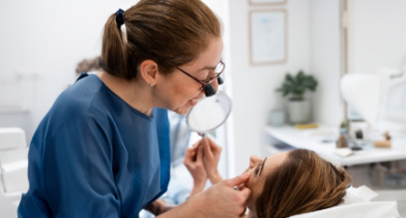
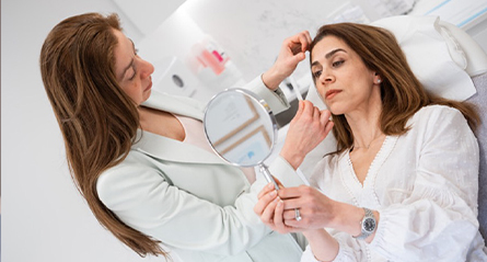





Before your consultation
Common questions, answered plainly.
What are the warning signs of a melanoma on the face?
Melanoma is the most serious of the common skin cancers, and early detection matters. The ABCDE guide is useful: Asymmetry, an irregular Border, uneven Colour, Diameter larger than about 6 mm, and Evolving or changing over time. On the face, watch for a new dark spot, a mole or freckle that is changing, or a slowly enlarging brown patch on the cheek, nose or ear. Arrange urgent GP assessment.
What should I do if I think I have a melanoma?
See your GP or dermatologist urgently rather than waiting to see whether the spot settles. The lesion will be examined, often with dermoscopy, and usually removed as an excision biopsy. Diagnosis is confirmed by histopathology, which reports the thickness and other features used for staging. A referral allows a Medicare rebate to be claimed for these procedures.
How is a facial melanoma treated after diagnosis?
Once histopathology confirms a melanoma, a wider local excision is usually planned around the biopsy site, with the margin based on the reported thickness. On the face this is balanced against the eyelid, nose, lip and ear, and reconstruction may use a local flap or a skin graft. For some melanomas a sentinel lymph node biopsy and staging investigations are discussed. Margins are checked by pathology and further surgery is sometimes needed.
What follow up happens after melanoma surgery?
Follow up is ongoing and usually involves scheduled reviews of the scar, the skin generally and the lymph nodes, with imaging or referral to a melanoma multidisciplinary team where indicated. Your schedule depends on the type, size, site and stage of the lesion. Continue regular skin checks with your GP or dermatologist, protect yourself from the sun, and report any new or changing spot promptly.