

- Home
- Procedures
- Face
- Skin Cancer
- Basal Cell Carcinoma (BCC)
Basal Cell Carcinoma (BCC)
Basal cell carcinoma (BCC) is a skin cancer that arises from the basal cells in the deepest layer of the epidermis. It is the most common skin cancer in Australia, and the face is the most common place to find one, particularly the nose, cheek, forehead, eyelid and ear. Long term ultraviolet exposure is the main contributing factor.
On the face a BCC typically presents as a small, pearly or translucent bump with fine blood vessels visible across it, or as a flat, slightly scaly patch that bleeds easily and scabs over. Some look like a persistent pimple that never quite settles, and others resemble a scar.
BCC grows slowly and rarely spreads to other parts of the body. Left untreated, however, it continues to extend locally, and on the face that can involve the eyelid margin, the nostril or the ear cartilage, which makes later repair more involved. If you have a spot on your face that fits this description, see your GP or dermatologist rather than watching it, and do not delay if a lesion is changing quickly or looks pigmented.
Signs to look out for
- A pearly, pink or skin coloured lump with small blood vessels on its surface.
- A spot that bleeds after minor contact such as shaving or washing, then scabs and bleeds again.
- A flat, pale or slightly scaly patch that slowly widens and can resemble a scar.
- A sore on the nose, ear or eyelid that partly heals then breaks down again.
- Loss of eyelashes or a change in the shape of the eyelid margin.
- A raised, rolled edge around a central dip or ulcer.
How a facial BCC is diagnosed
Your lesion is examined with dermoscopy, and the surrounding facial skin is assessed for laxity, natural creases and previous surgery, all of which affect how a wound can be closed.
Diagnosis is confirmed by biopsy and histopathology. The pathologist reports the BCC subtype, which matters on the face: nodular and superficial BCCs are usually well defined, while infiltrative and morphoeic subtypes send strands of tumour beyond the visible edge and need a wider margin or a different approach. The report also states whether the margins of an excision are clear.
How it is treated
Surgical excision is the usual treatment for a facial BCC. Some superficial lesions can be managed non-surgically by a dermatologist. Where a BCC sits on the eyelid, nose or ear, is recurrent, or has an infiltrative subtype, Mohs surgery may be recommended.
- Planning. The lesion is measured and marked, the margin is set for that subtype and site, and the reconstruction is discussed beforehand. Most facial BCC excisions are day procedures under local anaesthetic.
- Excision. Dr Maryam removes the BCC with a cuff of surrounding tissue and orientates the specimen for the pathologist. In Mohs cases the Mohs surgeon performs the excision and checks the margins during the procedure, and Dr Maryam performs the reconstruction.
- Margin checking. For a standard excision the pathologist reports whether tumour reaches any margin. This takes several days, and further surgery is sometimes needed.
- Reconstruction. The wound is closed in the way that suits the site: direct closure along a natural line, a local flap that borrows adjacent skin of similar colour and thickness, or a skin graft. Eyelid, nose and ear repairs may need cartilage support or a staged procedure.
Recovery and follow up
Most people are home the same day. Timeframes vary with the size of the excision, the site and the type of reconstruction.
- Oedema and bruising are common in the first week or two, particularly around the eyes and nose.
- Sutures on the face are generally removed within about five to ten days, and staged flaps involve a second visit to theatre some weeks later.
- Scars are pink and firm at first and usually soften over roughly six to twelve months.
- Your histopathology is reviewed with you at follow up, including whether any further excision is advised.
- Ongoing skin checks with your GP or dermatologist and daily sun protection remain important, because a further BCC elsewhere is common.
Your care with Dr Maryam
Dr Maryam Seyedabadi, Specialist Plastic Surgeon, MBBS FRACS, consults at 2c Heeley Street, Paddington in Sydney and operates at The Skin Hospital in Darlinghurst, St Luke’s Hospital in Potts Point, East Sydney Private Hospital, Sydney Day Hospital and Double Bay Day Hospital. A referral from your GP or dermatologist allows a Medicare rebate to be claimed for these procedures.
What surgery involves and how the area looks afterwards depend on the type, size, site and stage of the lesion, and on what the pathology report shows. These points are discussed with you before you decide. To arrange an appointment, call 1300 067 668.


What actually happens
Your Journey, Step by Step
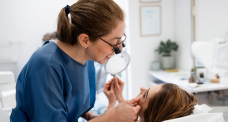
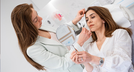





Before your consultation
Common questions, answered plainly.
What is a basal cell carcinoma on the face?
Basal cell carcinoma, or BCC, is the most common form of skin cancer. It arises from cells in the lower layer of the epidermis, grows slowly and rarely spreads to other parts of the body, although it can gradually invade nearby tissue. On the face it often presents as a pearly or shiny bump, a flat scaly patch, or a small sore that bleeds easily and keeps returning.
How is a BCC on the face diagnosed?
Your GP or dermatologist examines the spot, usually with dermoscopy, and takes a biopsy under local anaesthetic. Histopathology confirms whether it is a BCC and reports the subtype, since some subtypes extend further beneath the surface than they appear. That report guides the margin planned around the lesion. A referral allows a Medicare rebate to be claimed for these procedures.
How is a facial BCC treated surgically?
The usual treatment is excision with a planned margin of surrounding skin, followed by reconstruction using direct closure, a local flap or a skin graft. For a BCC on the eyelid, nose or ear, or for a recurrent lesion, Mohs surgery may be recommended, and the Mohs surgeon performs that excision while Dr Seyedabadi carries out the reconstruction. Margins are checked by pathology and further surgery is sometimes needed.
Can a BCC come back on the face after surgery?
Recurrence is possible, particularly with certain subtypes, larger lesions, and sites such as the nose, eyelid and ear where the margin available is limited. Having had one BCC also raises the chance of developing another elsewhere, so outcomes vary with the type, size, site and stage of the lesion. Ongoing skin checks with your GP or dermatologist and careful sun protection are recommended.