

- Home
- Procedures
- Body
- Skin Cancer
- Melanoma
Melanoma
Melanoma is a cancer that begins in the pigment producing cells of the skin. On the body it is found most often on the back and shoulders in men and on the lower legs in women, and it can present either as a new spot or as a change in a mole you already have.
Melanoma on the trunk and limbs is frequently noticed late for a simple reason: you cannot see most of your own back. A spot between the shoulder blades can change for months without being seen, while a lesion on the shin or calf may be dismissed as a mole that has always been there. Nodular melanoma is worth knowing about, because it grows as a firm raised lump, sometimes pink or red rather than brown, and it can change over weeks rather than years.
Unlike basal cell and squamous cell carcinoma, melanoma can spread beyond the skin, so assessment covers both the lesion and the lymph nodes that drain that region. Outcomes depend on the type, size, site and stage of the lesion.
Signs to look out for
- Asymmetry: one half of the spot does not match the other.
- Border: uneven, notched or poorly defined edges.
- Colour: several colours in the one spot, or an area that has darkened or lost colour.
- Diameter: wider than about 6mm, or steadily widening.
- Evolving: any change in size, shape, colour, height or surface, including itch, bleeding or crusting.
- Region specific signs: a lump on the back or shoulder that catches on clothing or bleeds, a spot on the lower leg that someone else notices has changed, or a firm raised lesion that has grown over a few weeks and needs review without delay.
How melanoma is diagnosed
A suspicious lesion is removed by excision biopsy, usually under local anaesthetic, and examined by histopathology. The report sets out the melanoma subtype, the Breslow thickness in millimetres, meaning how deeply the tumour has grown, whether the surface is ulcerated, and the mitotic rate. These details determine the margin required and whether further staging applies.
Early detection matters, and the back and legs are areas where a mirror or a second pair of eyes helps. If a spot fits any of the signs above, see your GP or dermatologist promptly rather than watching it for a few more months.
How it is treated
Treatment on the trunk and limbs balances an adequate margin against the closure that region will tolerate.
- Wider local excision around the biopsy scar, with the margin of normal skin guided by the Breslow thickness.
- Closure planned for the site: the trunk often has enough laxity for direct closure, although the wound sits under tension from shoulder and torso movement, while thin skin over the shin may need a skin graft or a local flap.
- Consideration of sentinel lymph node biopsy for some melanomas, sampling the armpit for back and shoulder lesions or the groin for leg lesions, commonly at the same operation as the wider excision.
- Staging scans, multidisciplinary team discussion or medical oncology referral where the pathology or nodal result indicates it.
Recovery and follow up
Recovery on the body is usually driven by movement and swelling rather than by appearance.
- Back and shoulder wounds are commonly protected from heavy lifting, stretching and gym work for around four to six weeks, as advised for your repair.
- Lower leg wounds swell, so elevation, walking little and often, and sometimes compression are used, and healing may take several weeks or longer.
- Scars on the trunk and limbs tend to stretch and stay firm for months, and often remain more noticeable than facial scars.
- Full skin checks continue long term, because a further melanoma or a recurrence is possible and follow up is ongoing.
- Sun protection every day, with clothing that covers the back and shoulders, a broad brimmed hat, broad spectrum sunscreen and shade.
Your care with Dr Maryam
Dr Maryam Seyedabadi is a Specialist Plastic Surgeon, MBBS FRACS, AHPRA MED0001341746, consulting at 2c Heeley Street, Paddington in Sydney. She operates at St Luke’s Hospital in Potts Point, East Sydney Private Hospital, Sydney Day Hospital, The Skin Hospital in Darlinghurst and Double Bay Day Hospital. A referral from your GP or dermatologist allows a Medicare rebate to be claimed for these procedures.
Your consultation covers the pathology findings, the margin planned, how the wound on that part of your body is likely to be closed and to scar, and the risks of the operation. What can be achieved depends on the type, size, site and stage of the lesion. To arrange an appointment, call 1300 067 668.


What actually happens
Your Journey, Step by Step
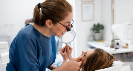
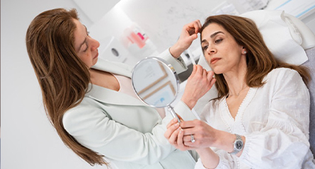





Before your consultation
Common questions, answered plainly.
How do I tell if a mole on my back or leg is a melanoma?
Melanoma is the most serious of the common skin cancers and often appears on the back, chest and legs. The ABCDE guide is a useful prompt: Asymmetry, irregular Border, uneven Colour, Diameter over about 6 mm, and Evolving over time. A spot that looks different from your others, itches or bleeds also needs checking. You cannot tell from appearance alone, so see your GP promptly.
How is a melanoma on the body diagnosed?
An excision biopsy of the whole lesion is the usual first step, done under local anaesthetic by your GP, dermatologist or surgeon. Histopathology confirms the diagnosis and reports Breslow thickness, ulceration and mitotic rate, which are used for staging. These findings guide the wider excision margin recommended and whether further tests apply. A referral allows a Medicare rebate to be claimed for these procedures.
What does wide local excision on the trunk or a limb involve?
A margin of normal skin is removed around the original biopsy scar, with the width guided by the thickness reported on pathology. Many wounds close directly, while larger excisions on the shin, shoulder or back may need a skin graft or a local flap. A sentinel lymph node biopsy is sometimes performed at the same time. Margins are reported by pathology and further surgery is occasionally needed.
What is a sentinel lymph node biopsy for?
It is a staging test, offered for some melanomas depending on thickness and other pathology features. A tracer identifies the first lymph node or nodes draining the melanoma site, and those nodes are removed and examined. The result helps clarify the stage and guides whether ongoing treatment or surveillance with a melanoma team is advised. Outcomes depend on the type, size, site and stage of the lesion.